Duke Eye Center may have solved a decades-old mystery in ophthalmology that could pave the way to treating a rare, incurable form of blindness.
People with Best vitelliform macular degeneration, or Best disease, often keep seeing clearly despite a large, egg yolk-like deposit forming in the macula, the part of the retina used for reading and recognizing faces.
The buildup pushes the eye’s light-sensing photoreceptors away from the support cells that keep them alive, a separation that, in theory, should severely damage eyesight. Yet patients retain near-normal vision for years, even decades.
Now scientists say they finally understand why.
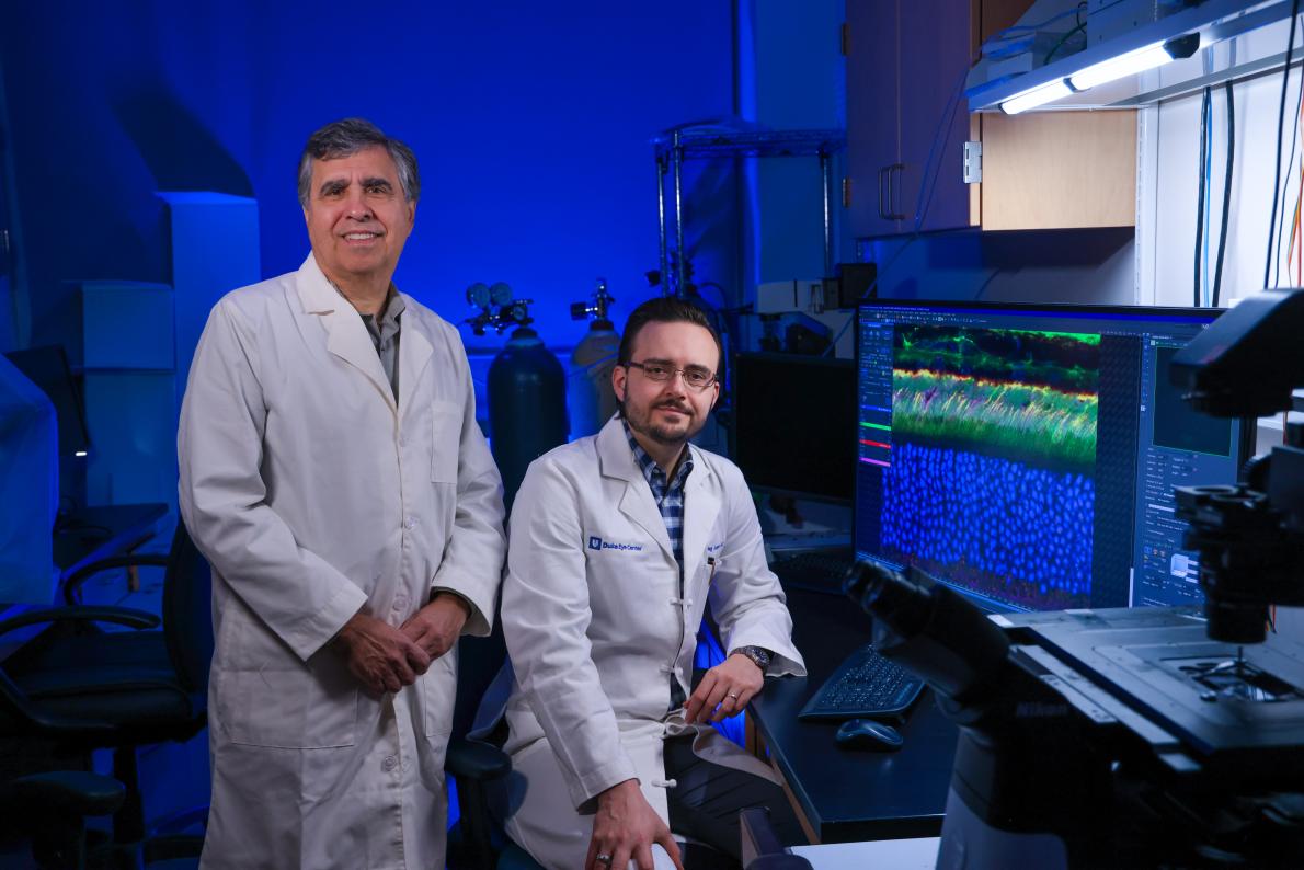
“This has been a persistent conundrum in ophthalmology,” said Vadim Y. Arshavsky, PhD, a professor of ophthalmology at Duke University School of Medicine who studies vitreoretinal diseases at Duke Eye Center. “Using electron microscopy and three-dimensional electron tomography, we were able to uncover how this is possible.”
Working alongside Tylor R. Lewis, PhD, an assistant professor at the University of Alabama at Birmingham, and Oleg Alekseev, MD, PhD, an assistant professor of ophthalmology at Duke, Arshavsky’s team uncovered a surprise hidden rescue system inside the eye.
When key retinal layers pull apart, support cells known as retinal pigment epithelium (RPE) send out ultra-thin, thread-like extensions. These tiny bridges stretch across the widening gap, allowing the cells to keep doing the daily cleanup and renewal work that light-sensing cells need to survive and preserve vision.
Researchers discovered the structures while studying mice engineered to lack a gene called ADAM9. Best disease is caused by multiple gene mutations. The mice developed the same kind of enlarged space seen in human Best disease, yet their light-sensing cells kept functioning far longer than expected.
The reason, according to the study in the Journal of Clinical Investigation, was the presence of the tiny extensions called pseudopods.
“These protrusions are very fine and difficult to detect even with powerful microscopes, which is why they had gone undetected until now,” said Arshavsky, senior study author.
Implications for treatment

The finding resolves a long-standing contradiction in ophthalmology. For years, scientists believed photoreceptors and the RPE had to remain tightly pressed together for vision to work. Pull them apart, and the renewal process that sustains vision should fail.
This discovery helps explain how vision can stay surprisingly strong even when the eye’s structure is disrupted. It could also inform new gene therapies now being tested for Best disease and similar conditions.
“Understanding how the retina compensates on its own could help with the design of treatments that support or even strengthen this natural back up system,” said Lewis, the study's lead author who conducted the work as a postdoctoral researcher at Duke Eye Center.
Microscopic rescue mission
But the rescue wasn’t permanent. Over time, perhaps when the deposits grow too large, the pseudopods dwindle. The mice’s photoreceptors began to degenerate, leading to vision loss.
“The RPE pseudopods we observed are about 5 to 10 microns wide, roughly 10 times thinner than human hair,” Lewis said. “By comparison, vitelliform lesions in patients measure about 1 millimeter, or 1,000 microns. That means hundreds of these slender extensions would likely need to form within a single lesion to adequately support the photoreceptor cells beneath it.”
The structures also appear to be highly dynamic, constantly forming and retracting as they work to sustain large numbers of photoreceptors.
The same mechanism could also play a role in other retinal conditions marked by subretinal deposits that force cells apart, including certain forms of macular degeneration.
More studies are needed to learn exactly how these pseudopods form and whether they appear in other human eye diseases. But the work offers a new view of a problem that has long defied explanation.
This work was funded by the National Institutes of Health, Duke University Physician-Scientist Strong Start Award, and Research to Prevent Blindness.
Additional authors include Carson M. Castillo, Sebastien Phan, Camilla R. Shores, Kylie K. Hayase, Keun-Young Kim, Mark H. Ellisman, Oleg Alekseev, and Marie E. Burns.