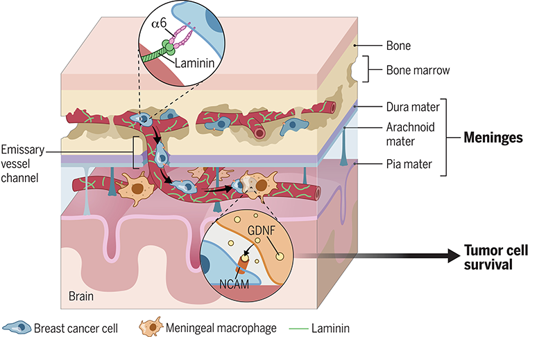
Once cancer gets inside the inner membranes that protect the brain and spinal cord (the leptomeninges), the disease can circulate in the cerebrospinal fluid and spread throughout the central nervous system. The outcome for patients is devastating, with a median survival time of less than six months.
That’s why clinician-scientist Dorothy Sipkins, MD, PhD, and her team at Duke University School of Medicine have kept pushing for nearly 10 years to find out how cancer cells can get inside this vital compartment.
Now they have discovered a previously unknown shortcut that some breast cancer cells use to metastasize to the leptomeninges, as well as clues that suggest how to block this path. Their study was published June 21, 2024, in the journal Science, with an accompanying commentary.
Sipkins, an associate professor of medicine, and her team showed in mice that breast cancer cells first infiltrated the bone marrow of the skull, then traveled via “emissary blood vessels.” These vessels begin in the bone marrow, pass through normal openings or “windows” in the skull, then become part of the leptomeninges.
“These tumor cells grab onto the outside of these blood vessels and basically shimmy down the vessel like a fireman sliding down a fireman’s pole,” said Sipkins, a member of Duke Cancer Institute.

The work suggests a potential way to prevent cancer cells from taking this shortcut. “The cells in our study expressed a receptor called integrin alpha6 that they use to grab onto a specific protein that encases these blood vessels. Our evidence suggests that this protein, laminin, greases the fireman’s pole,” she said.
Sipkins hopes these findings will lead to a way to identify which women with breast-to-bone metastases are at highest risk for leptomeningeal disease. “If we can identify those patients by the presence of this marker integrin alpha6, could we prevent the spread?” she said. “And then for patients who already have the spread of the disease, what can we do for them?”
“We haven’t moved the dial in many years in terms of improving treatments for breast cancer patients with leptomeningeal metastasis,” Sipkins said. The disease is rare but is becoming more common as people live longer with breast cancer. “The symptoms that patients experience from this are debilitating. It's a terrible complication,” she said.
After the team found out how breast cancer cells get inside the leptomeninges, they pushed further to figure out how the tumor cells survive there.
Using their tool of choice — confocal microscopy — the researchers peered into the meninges and saw that the cancers cells stuck close to a particular type of immune system cell called macrophages. Then the tumor cells stimulated the macrophages to secrete a protein (glial-derived neurotrophic factor) that is normally used to protect neurons (brain cells).
“We demonstrated that in our mouse models, glial-derived neurotrophic factor from meningeal macrophages was important for breast cancer cells to survive and proliferate, and to be successful in the meningeal microenvironment,” Sipkins said. She hopes to use this knowledge to develop new treatments.
Sipkins praised her team’s dogged determination to find these answers. For example, post-doctoral associate Danhui Ma, PhD, spent countless hours learning new microscopy techniques and conducting final experiments, Sipkins said.
The initial work that went into this study would not have been possible without a philanthropic gift from David Wells, Sipkins said. Wells’ wife Leslie passed away after a brief battle with leptomeningeal disease, which she fought with the help of the Duke Center for Brain and Spine Metastasis. He made the gift in her honor.
As a clinician, Sipkins treats patients with leukemia, and that is her main research focus. But she began studying breast cancer as well several years ago because it often spreads to the bone and bone marrow. This new study was inspired by some of her previous work in leukemia, in which she discovered that acute lymphoblastic leukemia cells can use this “fireman’s pole” method to spread to the leptomeninges.