
First Encounters with the Inner Brain

Standing in a brightly lit teaching lab in the Mary Duke Biddle Trent Semans Center for Health Education, Leonard White pops the lid off a bucket, reaches in with gloved hands, and carefully pulls out a three-pound human brain, roughly the size of a cantaloupe.
The Duke neurology professor says today is a big day for the 129 first-year medical students in his Brain and Behavior class.
In their first six months of medical school, the students have mostly examined the brain from the outside. They’ve run their fingers over its wrinkled walnut-like surface and traced the deep fissures that separate its lobes.
But today, White says, these future medical doctors will peer inside and get a firsthand glimpse of what lies beneath the surface.
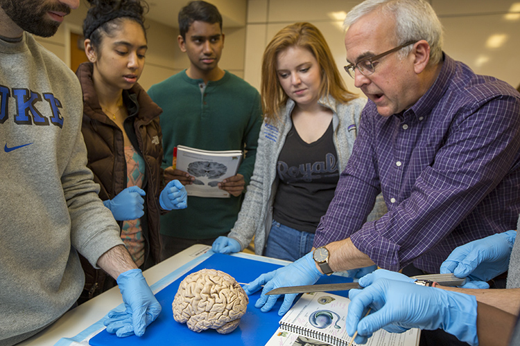
As the students file into the room they make their way among rows of tables laid out with brains. Some rest intact in plastic buckets. Others are sliced in cross section as they might see on a CT scan, and arranged on metal trays.
After everyone puts on gloves and finds their assigned station they get to work. Standing elbow to elbow in small groups, the students flip open their lab manuals and take turns reading out the names of the features they’re supposed to identify.
“OK so can we find the caudate-putamen and the accumbens?” one student says, glancing up from the page toward the brain.
They’re looking for masses of gray matter deep inside the brain that facilitate movement. This is where brains go wrong for people with diseases like Parkinson’s or Huntington’s who gradually lose the ability to do things like walk gracefully and maintain their balance.
As the students handle the slippery brain slabs they work together to get their bearings. “Which is the front and which is the back?” one student asks. “This is definitely posterior,” says another.
Covering the brain like an orange peel is a layer of gray cells, just a few millimeters thick.
Underneath this outer layer and filling nearly half the brain is the inner white matter. It contains a labyrinthine network of threadlike nerve fibers that connect neurons and carry signals between them -- the brain’s information superhighway.
A team of students locates a c-shaped cavity in the center of a piece of the brain. “Here’s the lateral ventricle,” one students says. Then, using that as a landmark, “shouldn’t the caudate be right here?”
Another student uses a wooden probe to point to alternating gray and white strands. “So these stripes are the striatum, right?” she says, and a chorus of “Oh!” erupts from the group.
White and other instructors circulate around the room, checking their progress. White stops at one table where the students are examining a brain. He calls their attention to a fist-sized structure tucked beneath its base. In cross section the dense folds of white and gray matter look like a split head of crinkly-leafed cabbage.
“This is a very beautiful cerebellum,” White says.
Brain and Behavior is one of four basic science courses required of all first-year medical students at Duke. It’s a grueling curriculum, condensing what many medical schools take 18 months or more to teach into one year to provide students more opportunities to pursue their individual interests through research and electives in years three and four. Classes run five days a week. When they’re not in the lab or lecture hall, the students are watching video tutorials and going over the day’s assigned readings or studying for exams.
The course is also an opportunity to learn from educators like White. An associate professor of neurology, he has been honored four times with the Duke Health Golden Apple Award, the most prestigious teaching award presented by the Duke University School of Medicine student body. He is also a co-author and co-editor of a major neuroscience textbook.
As the course’s primary director, White is captivated by the brain. The most complex organ in the human body -- as it is often described -- contains a staggering 100 billion neurons, nearly the number of stars in the Milky Way.
He remembers the first time he held one, while working on a master’s degree in physiology at Oral Roberts University. It was 1986, the same year he married Heidi, now his wife of nearly 33 years. And in both cases, he says, it was “love at first sight.”
White has since dedicated himself to a career in neuroscience, and now more than three decades later he’s guiding other students through a similar rite of passage.
“We’re trying to make strong connections between anatomy and function so that students can take that knowledge into the clinical realm,” White said.
The students don’t know who donated the brains. But from the labels on the buckets they learn that one woman died at age 69 of a heart attack, another at 94 from the flu. At least half a dozen people lost battles with cancer.
At a nearby table, a group of students study a brain, hesitating before making their first incision in the organ that once produced the person’s every thought, memory, emotion and action.
Their task is to try to identify the same deep gray matter structures as before, but using different planes of dissection. The idea is to help students make sense of cross sections of the brain from any angle, the same views of the brain they might one day need to interpret in a patient’s CT scan or MRI. As they do, they’re recalibrating their 3-D mental map of the brain’s interior based on what they see in front of them.
No two brains are exactly alike, White says. “We all have the same basic parts, but their size, shape, and position relative to one another can be remarkably different from one person to the next.”
From one brain the students learn to recognize the telltale scarring from chronic hypertension. From another, they see what damage a stroke can do.
One woman has a marble-sized hole in her cortex. A stroke at some point in her life must have cut off blood flow to that part of the brain, and without oxygen the brain cells in the area died, leaving a void.
After dissection is done for the day the students begin cleaning up and putting things away. White turns his attention to a brain lying in a tray, and says, “I’m looking for a beautiful amygdala. Can you point to it?” After a second of reflection a student touches a gloved finger to an almond-shaped structure. “Yep, that’s exactly it.”
Robin Smith is senior science writer in the office of Duke University Communications, where she covers the life and physical sciences across campus.
Julie Schoonmaker is the video manager and Jared Lazarus is a photojournalist with the office of Duke University Communications, where they enjoy bringing stories about research, discovery and learning to life.