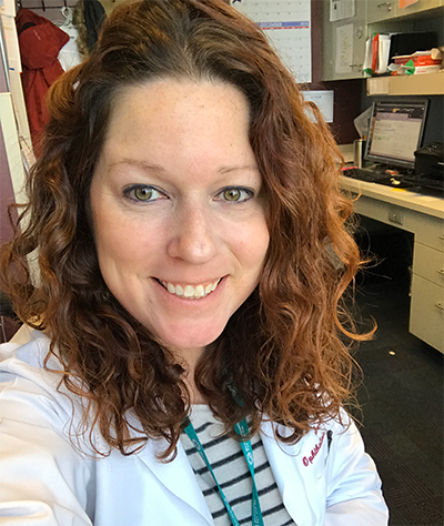
After a bachelor’s degree in visual journalism and 15 years working as a freelance photographer shooting portraits, events, sports, and food and beverages, Kathleen Warren never imagined herself in a medical field. But in 2013, a friend from college encouraged her to join him at the New England Eye Center as an ophthalmic photographer. When she investigated it, she liked the possibilities.
“If you think about it, our eyes are our camera lenses to the world,” Warren said. “The way that we perceive light, the way that we see things, the signals that we get, our color palettes, all of it, are basically like a camera.”
She accepted the photographer job at the New England Eye Center in Boston, Massachusetts, where for more than three years she shadowed other ophthalmic photographers, learning on the job. “My brain was like a sponge,” she said. After three more years, she moved to the Massachusetts Eye and Ear Infirmary for six years before joining the Duke Eye Center and Duke University Hospital in 2019, where she works on a team of 12 ophthalmic photographers.
“It’s like I have 11 brothers and sisters,” she said. “We work well together. We service the whole hospital when it comes to eyes. If you need any kind of images: fundus photos, anterior segment of your cornea, your optic nerves. Anything that has to do with the anatomy or physiology of the eye, we are your team.”

Warren counts all her previous photography experience as preparing her for her career in ophthalmic photography. “Understanding the aesthetics of the cameras is important,” she said. Though she leaves diagnoses to the clinicians, Warren said that she has learned to spot something that looks unusual that she needs to explore with additional images. “It's ultimately getting the best images for the patient, so the doctor has all the necessary information to diagnose them,” she said.
“It's so rewarding to be able to help people with their vision,” she said. “I trained under some of the best surgeons and retina doctors, worked in the OR, traveled for educational conferences, and have learned so much in this field that I wouldn't trade it for anything.”
Warren loves the patient-focused atmosphere at Duke. “We take the extra time to notice our patients and to be there with them,” she said. “We never make them feel rushed. I've cried with patients, held their hands, and been there as an extra blanket of comfort.”
She holds two technical medical credentials. She and other members of the team are certified phlebotomists, a skill they use to perform retinal angiography, which requires injecting dye into a vein to take pictures of the circulation within the eye’s blood vessels. She’s also certified in optical coherence tomography, which is a non-invasive imaging technique that uses light to produce cross-sectional images of the retina.
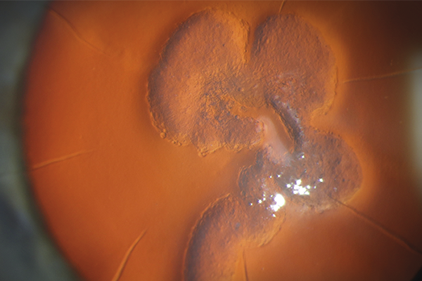
In 2022, Warren won top 10 honors in an international competition for images taken with a Haag-Streit slit lamp camera, which uses a magnifying microscope and a bright light to illuminate the eye to examine structures like the cornea, the iris, the vitreous, and the retina. She received third place among 168 images submitted by participants from 20 countries for her image of a case of epithelial ingrowth (a rare but serious complication that can occur in the eye after trauma or surgery).
“The Haag-Streit is the hardest camera to master, in my opinion” Warren said, “If it’s not lit properly, you can really miss the mark on things.”
Warren radiates high energy, and she needs it. In addition to her full-time work at Duke, she has a storefront, “Uprooted,” where she sells her creative photography and art. She serves as editor-in-chief of the Journal of Ophthalmic Photography for the Ophthalmic Photographers’ Society, as well as chair of their scientific exhibit committee. In addition, she was recently accepted to graduate school at Tulane University, where she will pursue an online master’s degree in social work.
“I just consume myself with work a lot, but it's not really work,” Warren said. “It's just fun.”