

A discovery that began in a Duke laboratory is now being tested in Duke patients, offering a potential new approach to diagnosing cardiac sarcoidosis, which can be life-threatening and difficult to diagnose.
The work grew from a collaboration between researchers in the Department of Molecular Genetics and Microbiology and cardiologist-scientist Ravi Karra, MD, director of the Cardiac Sarcoid Program in Duke’s Sarcoid Center for Excellence.
Their shared interest in macrophages — immune cells that play a central role in inflammation — led to a finding that has advanced from tissue studies to a Duke-led clinical trial.
Identifying granulomas in heart tissue

The Tobin Lab, led by David Tobin, PhD, studies granuloma biology in diseases including tuberculosis, while Karra treats patients with cardiac sarcoidosis. In a recent paper in the Journal of Cardiac Failure, Charlie Pyle, PharmD, PhD, a senior research associate in the Tobin Lab, and colleagues showed that tilmanocept, an FDA-approved imaging agent, can identify granulomas in heart tissue from patients with sarcoidosis.
Granulomas are organized clusters of immune cells that form during inflammation and are a hallmark of sarcoidosis. When they develop in the heart, they can cause serious complications, including abnormal heart rhythms and heart failure.
“We uncovered a new way to identify lesions in the disease sarcoidosis,” Pyle said. “We discovered that the immune cells in sarcoidosis granulomas, called macrophages, have high amounts of a protein that the imaging probe recognizes.”
The collaboration
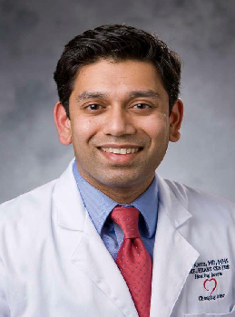
The collaboration began when Pyle was helping members of Karra’s group with tissue staining. Pyle and Mohamed El-Ganainy, then a trainee in the group, noticed high levels of MRC1, also known as CD206, on macrophages in sarcoidosis tissue.
Pyle was familiar with tilmanocept because his graduate school adviser had studied MRC1 as the imaging agent’s target. He suggested testing whether tilmanocept could bind to macrophages within sarcoidosis granulomas.
Experiments showed a significant interaction between MRC1 and tilmanocept in these granulomas.
The researchers found substantially higher levels of CD206-positive macrophages in cardiac sarcoidosis tissue than in tissue from patients with other cardiac conditions. A fluorescent version of tilmanocept also accumulated within granulomas, suggesting the imaging agent could distinguish sarcoidosis lesions from surrounding tissue.
A challenging disease
The finding could address a significant challenge in diagnosing cardiac sarcoidosis since existing noninvasive imaging can detect inflammation or scarring but may not be specific to sarcoidosis.
“Sarcoidosis is a challenging disease because we have really good treatment options, but it can be hard to diagnose,” Karra said. “When it gets into places like the heart, we don’t have reliable methods to diagnose it for sure.”
The laboratory findings have now moved into a Duke-led clinical trial, in which Karra is testing radiolabeled tilmanocept in Duke patients to determine whether the imaging agent can identify cardiac sarcoidosis in living patients.
“This could lead to a new way to diagnose sarcoidosis in patients that is safer and more reliable,” Pyle said.
For Karra, the potential benefit is greater certainty in deciding who needs treatment.
Benefiting more patients
“Right now, because sarcoidosis — especially in the heart — can be very dangerous, patients often need to be treated based on their probability of being affected,” he said. “A better diagnostic test could help us better identify patients who might benefit from treatment and avoid treating patients who may look like they have sarcoidosis but actually have something else.”
The researchers continue to study how macrophage markers influence granuloma formation and which methods of administering tilmanocept might produce the best imaging results.
The project demonstrates how collaboration across Duke departments can move a basic-science discovery toward patient care — from an observation about immune cells in diseased tissue to a clinical trial testing the finding in Duke patients.