Researchers at the Duke Eye Center have made an important – and surprising – discovery in human eye anatomy that has gone unnoticed for decades. Using enhanced imaging technology, they identified a previously unknown part of photoreceptor cells in the retina. Their finding, published in Communications Biology, challenges long-standing assumptions about the retina and opens new avenues for understanding vision and genetic eye diseases.
Most of what researchers know about retinal anatomy has come from exhaustive studies using electron microscopy in the 1960s and 1970s. Eventually, no new major discoveries were being made in the anatomy of the retina, so that chapter of research closed.
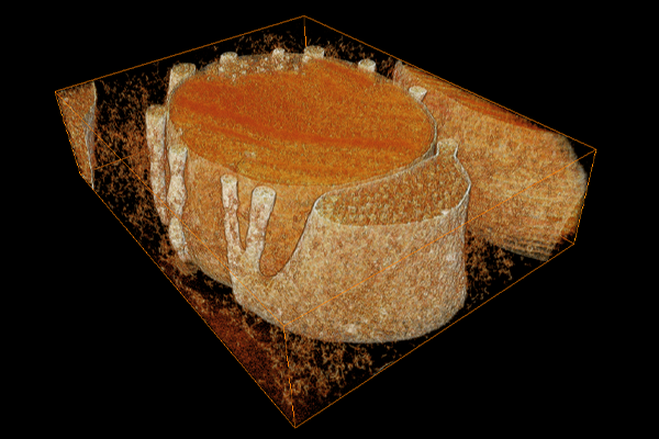
But technology kept advancing. Oleg Alekseev, MD, PhD, assistant professor of ophthalmology, and Vadim Arshavsky, PhD, Helena Rubinstein Foundation Distinguished Professor of Ophthalmology, decided to use three-dimensional electron tomography, which creates a 3D map of an object, to look at retinas with unprecedented resolution. “We aimed to take a look at some very small structures,” Alekseev said, “but we found something pretty major.”
Inside the retina are photoreceptor cells, commonly known as rods and cones. Photoreceptor cells convert light signals into electrical messages that the brain decodes and processes into visual images. Alekseev, Arshavsky, and team found that each rod photoreceptor cell contains a large, mechanically reinforced protrusion, which they’ve termed the accessory inner segment (aIS).
“The function isn’t known yet,” Alekseev said, “but we presume it serves as a physical support to hold the photoreceptor.” Photoreceptors are long and narrow. The researchers think that the aIS holds everything together and stable, preventing the photoreceptors from collapsing and flopping.
Over 300 known genes make photoreceptors functional. A mutation in any one of those genes can make the photoreceptor unable to work properly and can lead to irreversible blindness. Genetic testing helps solve only around 70% of cases, but this discovery opens the door to studying additional previously overlooked genes that may be involved in forming and maintaining the aIS.
“We need to explore this structure further,” Alekseev said. “It’s very likely that some diseases are caused by defects in the aIS, and now we have the opportunity to explore those diseases, improve our ability to diagnose patients, and start thinking about treatments.”
The aIS appears to be a uniquely human structure, which could explain some of the discrepancies between human diseases in the eye and mouse models of these diseases. For example, humans with Usher syndrome experience both blindness and deafness. In mouse models, though, introducing the exact same mutations in the exact same genes produces mice that are deaf, but not blind.
“We speculate,” said Arshavsky, “that some proteins in humans may be functioning within the aIS structure, and whenever you have a discrepancy between a human and mouse phenotype of some gene mutations, this structure comes into play.”
Now that aIS has been discovered, the next step is to understand the molecular composition of its unique parts and see what questions they can answer about human diseases in the eye.